


Prepare for the experiment by putting on gloves, wearing a lab coat, and gathering materials. See Things You'll Need below for reference. Grab a slide and rinse it with soap and water. Use Bibulous paper to dry the slide. Use a black sharpie to draw a circle on the bottom of the slide in order to identify the area for which bacteria will be smeared. Important laboratory safety-wear consists of goggles, closed toed shoes, gloves, and lab coat.

Flame the inoculating loop and wait 10 seconds before placing the loop into the pipette of bacteria. Make a smear of bacteria on the slide. Flame the loop again and place loop on the drying rack. Flaming the loop ensures any bacteria that may have resided on the loop prior to using it is killed off.
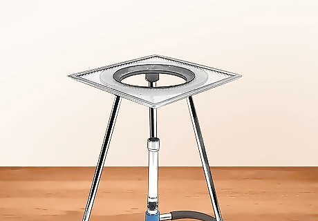
Set up a bunsen burner on your ring stand with the ring clamp and a wire gauze over it.
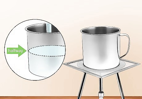
Fill the can with tap water until it is about half-full. Place the can on top of the wire gauze on the ring clamp.

Turn on the bunsen burner to heat/boil the water. Let steam start to appear before putting the slide on the can. Place the slide with smear on top of the can.
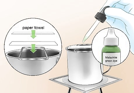
Place a piece of paper towel on top of the smear on the slide and drench the paper towel with the Malachite green dye.

Steam the smear over the water for 5 minutes. If the Malachite green evaporates, add more. Keep the paper wet. Use forceps to take the slide off when finished - it will be hot.
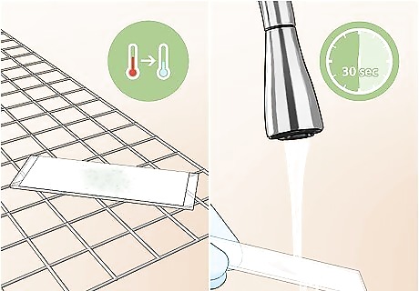
Allow the slide to cool, remove the paper, and rinse with water for 30 seconds.
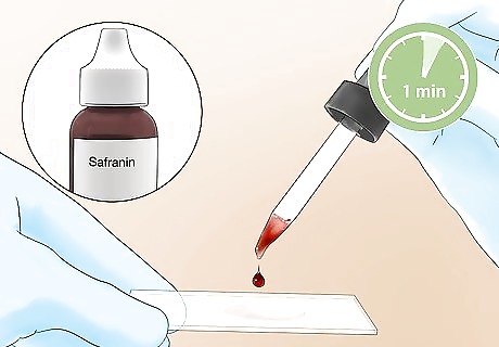
Add Safranin to the slide for 1 minute. The plastic beaker can be used to drain the excess dye.
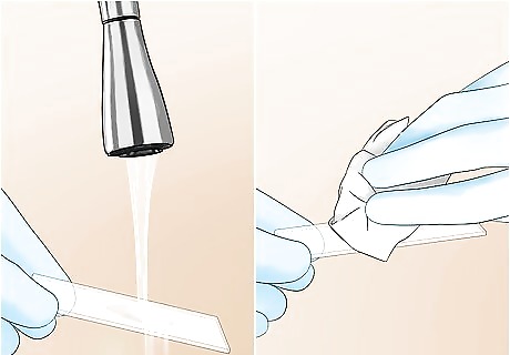
Rinse off the Safranin with water in order to remove excess dye. Blot the slide between sheets of Bibulous paper to dry. After the slide is dried it can be examined under a microscope.

View the slide. Place it on the stage. For this type of stain, the bacteria can only be viewed at the highest objective (100x), known as the oil immersion lens. This oil enhances resolving power and will allow the light to focus perfectly on the specimen.
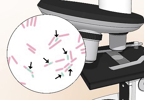
Examine the slide to see the endospore. The vegetative cell will appear red/pink, while the endospore itself is stained green. Spores may be located terminal, middle, or subterminal inside the vegetative cell.


Comments
0 comment